Parasites In Humans
Find The Nastiest Parasites In Humans
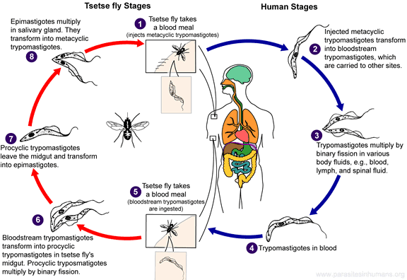
Trypanosoma Brucei - Sleeping SicknessSleeping sickness, African trypanosomiasis, is a deadly blood disease caused by two variates of Trypanosoma brucei and transmitted by tsetse fly. Trypanosoma brucei rhodesiense causes East African trypanosomiasis. 1000 new T. b. rhodesiense infections are reported to World Health Organization annually. Trypanosoma brucei gambiense causes West African trypanosomiasis (also known as Gambian sleeping sickness). More than 12 000 new infections are reported to the WHO each year. The two subspecies do not overlap in geographic distribution. They infect humans and tsetse flies (Glossina genus) in the woodlands, savannah and the dense vegetation between Kalahari and Sahara deserts. Less than 1 % of tsetse flies carry the parasite. T.b. rhodesiense is found in eastern and southeastern Africa. Over 95 % of the T.b. rhodesiense infections occur in Malawi, Tanzania, Uganda, and Zambia. T.b. gambiense is found predominately in central Africa and in some areas of West Africa. Over 95 % of the T.b. gambiense infections occur in Angola, Central African Republic, Chad, northern Uganda, Sudan, Republic of the Congo and Democratic Republic of the Congo. Trypanosoma brucei needs two hosts to live and reproduce. Its life cycle starts, when an infected tsetse fly bites human skin. While it is feeding on blood, metacyclic trypomastigotes are transmitted to the skin from the salivary glands of the fly. The parasites get into the bloodstream by entering lymphatic or blood vessels. They travel in different body fluids (such as blood, lymphatic or spinal fluid), transform into bloodstream trypomastigotes and multiply by binary fission. The disease can be spread by another tsetse fly that drinks the infected blood. Inside the fly the life cycle takes about three weeks. Ingested bloodstream trypomastigotes transform into procyclic trypomastigotes in the fly's midgut and multiply. They transform into epimastigotes, migrate to the salivary glands, then transform into metacyclic trypomastigotes and multiply once again by binary fission.
Trypanosoma brucei is not killed by the immune system because it has a glycoprotein (VSG) coating. The coating makes its cell membrane very thick and hard to recognize. It also changes frequently its structure to always keep ahead of the immune response. T. b. rhodesiense and T. b. gambiense are indistinguishable under a microscope. A trypomastigote is 14 to 33 µm long and has a tiny kinetoplast located at the posterior end, a centrally located nucleus, an undulating membrane, and a flagellum. Trypomastigotes are the only stage found in patients. Humans are the main host for T. b., but it is sometimes found in animals. East African trypanosomiasis is more acute and progresses quicker than West African trypanosomiasis. Symptoms can be minor in the beginning but usually become apparent within the first months of the infection. African trypanosomiasis has three symptomatic stages, the last one being the most dangerous eventually leading to death, if left untreated. 1. In 1–3 weeks after the bite a chancre (a red sore skin lesion) can develop on the bite area. 2. Several weeks or months later Trypanosoma parasites in the blood, spinal and lymphatic fluid (hemolymphatic stage) can cause:
3. The disease reaches its final stage when the parasites get through the blood-brain barrier entering the brain. The central nervous system involvement can occur as early as within a month in some cases. This meningoencephalitic stage (inflammation of the central nervous system) causes (in addition to the above second stage symptoms) some of the following symptoms:
Your health care provider does the diagnosis by examining blood, lymph node or tissue aspirates (fluid suction), bone marrow, chancre or cerebrospinal fluid under a microscope. Treatment is based on symptoms and laboratory results. The drug choice depends on the infecting species and the stage of infection. Pentamidine isethionate and suramin are usually used for treating the hemolymphatic stage of West and East African Trypanosomiasis, respectively. Melarsoprol is used for late disease with central nervous system involvement (infections by T.b. gambiense or T. b. rhodiense). Hospitalization for initial treatment is often necessary. Periodic follow-ups that include a spinal tap are required for two years. Tsetse flies are common only in rural Africa, not in urban cities. Areas of heavy infestation (endemic areas) should be avoided. Tsetse flies are attracted to bright colours, very dark colours and moving vehicles. Check for tsetse flies before entering the car. Tsetse flies are day biters. They can bite through thin clothing and the bite is usually very painful. Permethrin-impregnated clothing and use of DEET repellent (spray) can reduce the risk of getting bitten. Wear long-sleeved shirts and pants made of medium-weight material in neutral colours that blend in with the background environment. Avoid thickets because during the hottest period of the day tsetse fly rests in bushes. Use insect repellent. Even though insect repellents are not effective against flies, they are effective in preventing other insects (such as malaria-carrying Anopheles mosquitoes) from biting.
Also check out the videos.
|
Parasites In Humans Pictures And Videos Contact Info Privacy Policy Sitemap
© 2010–2015 parasitesinhumans.org


